Home
/ Sagittal Section Of Brain : anterior posterior inferior figure 2018 human brain ... : The fore brain is the main thinking part of the brain.
Sagittal Section Of Brain : anterior posterior inferior figure 2018 human brain ... : The fore brain is the main thinking part of the brain.
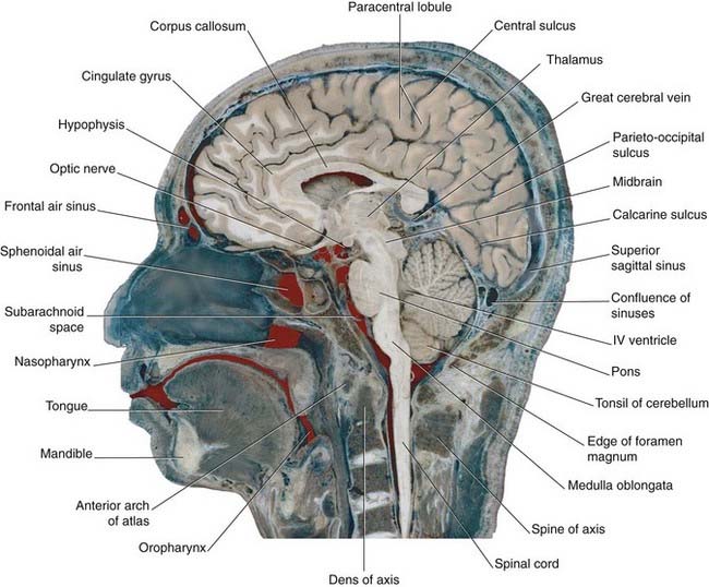
Sagittal Section Of Brain : anterior posterior inferior figure 2018 human brain ... : The fore brain is the main thinking part of the brain.. A midsagittal section of the brain shows the cerebrum, cerebellum and the brainstem. Sagittal section of human brain identify the structures in figure 20.18, sagittal section. Learn the functional areas of the brain in a midsagittal view, now at kenhub. Visible structures of the brain in sagittal section of brain. As per bridgeman art library v.
Brain, cerebral arteries, base of brain, diagram. Folds or 'bumps' on surface of cerebral cortex. Anomalies of midline structures of the brain are easily discernable on sagittal images; Diagrammatic images from grays (20th ed): The features indicated in the sagittal section of the right half of the human brain include body, corpus callosum;
Cerebral topography | Neupsy Key from neupsykey.com Use the diagram of the sagittal section to locate, observe and review the function of the following structures on the sheep brain sections The brain is divided into the prosencephalon. This exhibit depicts the normal sagittal section of the brain. Bundle of axons connecting r & l cerebral hemispheres. Each side of the brain controls the opposite side of the body. Contact us today to discover more about our available illustrations. The fore brain is the main thinking part of the brain. An mri was performed on a healthy subject, with several acquisitions with different weightings the vertical left menu provides reference images on coronal and sagittal views of the brain, with anatomical schemas based on a three dimensional (3d) model.
This study aimed to investigate.
Top suggestions for sagittal section of human brain. This is an online quiz called midsagittal section of brain. This high quality transparent png images is totally free on pngkit. Sagittal mri scan of brain of patient with chiari malformation.jpg 1,200 × 1,344; There is a printable worksheet available for download here so you can take the quiz with pen and paper. This picture also contains other parts such as interventricular foramen, monro, foramen monro, septum pellucidum, fornix, sulcus of corpus callosum, medial frontal gyrus, cingulate sulcus, cingulate gyrus, paracentral sulcus, cerebrum, brain in situ brain anatomy, brain sagittal section, brain medial view. Use the diagram of the sagittal section to locate, observe and review the function of the following structures on the sheep brain sections Identical twins at a gestational age of 15 weeks, shown in. A sagittal section through the human brain showing the structures of the left side. As per bridgeman art library v. Medical didactic anatomy illustration with the name and description of functional areas. Each side of the brain controls the opposite side of the body. This exhibit depicts the normal sagittal section of the brain.
Brain, cerebellum, sagittal section, gross. Map of the brain with all the basics. The term sagittal is derived from the latin word sagitta, meaning arrow. Medical didactic anatomy illustration with the name and description of functional areas. Sagittal section of brain the brain labeled anterior cerebral artery oculomotor nerve 3 internal carotid artery.
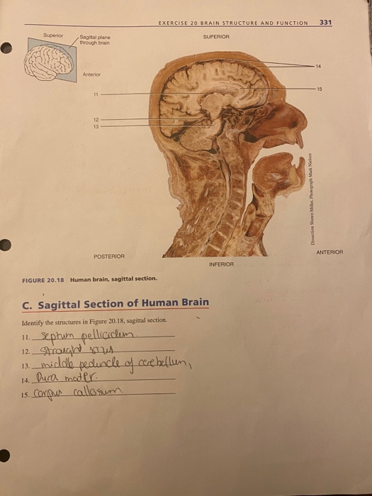
Solved: EXERCISE 20 BRAIN STRUCTURE AND FUNCTION 331 Super ... from media.cheggcdn.com Learn the functional areas of the brain in a midsagittal view, now at kenhub. There is no audio file. Use the diagram of the sagittal section to locate, observe and review the function of the following structures on the sheep brain sections The term sagittal is derived from the latin word sagitta, meaning arrow. Sagittal mri scan of brain of patient with chiari malformation.jpg 1,200 × 1,344; As per bridgeman art library v. This high quality transparent png images is totally free on pngkit. The brain is divided into the prosencephalon.
The ability to obtain sagittal sections counts as one of the merits of mri, although sagittal reconstructions may also be obtained from ct volume data.
Anomalies of midline structures of the brain are easily discernable on sagittal images; The brain is divided into the prosencephalon. This exhibit depicts the normal sagittal section of the brain. Diagrammatic images from grays (20th ed): This high quality transparent png images is totally free on pngkit. Vector 3d illustration isolated on white background. An mri was performed on a healthy subject, with several acquisitions with different weightings the vertical left menu provides reference images on coronal and sagittal views of the brain, with anatomical schemas based on a three dimensional (3d) model. An image of an arrow piercing a body and additional images. The features indicated in the sagittal section of the right half of the human brain include body, corpus callosum; Use the diagram of the sagittal section to locate, observe and review the function of the following structures on the sheep brain sections It has regions which receive sensory impulses from this is an important part of brain which controls the whole body. There is no audio file. Cut the brain in to two equal sagittal sections.
Sagittal section of human brain identify the structures in figure 20.18, sagittal section. Map of the brain with all the basics. There is no audio file. Brain, cerebellum, sagittal section, gross. The decline can be attributed to surgical effects, but the relative contributions of the stimulation parameters are not well understood.
Cerebral topography | Neupsy Key from neupsykey.com Visible structures of the brain in sagittal section of brain. Sagittal mri scan of brain of patient with chiari malformation.jpg 1,200 × 1,344; Diagrammatic images from grays (20th ed): Medical didactic anatomy illustration with the name and description of functional areas. Vector 3d illustration isolated on white background. Narrowing of the spinal canal by extradural. A midsagittal section of the brain shows the cerebrum, cerebellum and the brainstem. Sectional planes of the brain.
Use the diagram of the sagittal section to locate, observe and review the function of the following structures on the sheep brain sections
Map of the brain with all the basics. Any damage or injury to the sagittal section will affect the whole body. The fore brain is the main thinking part of the brain. Section of brain, by capieux, 1792. This study aimed to investigate. Identical twins at a gestational age of 15 weeks, shown in. A midsagittal section of the brain shows the cerebrum, cerebellum and the brainstem. The features indicated in the sagittal section of the right half of the human brain include body, corpus callosum; Sagittal section of human brain identify the structures in figure 20.18, sagittal section. Sagittal mri scan of brain of patient with chiari malformation.jpg 1,200 × 1,344; Use the diagram of the sagittal section to locate, observe and review the function of the following structures on the sheep brain sections Folds or 'bumps' on surface of cerebral cortex. Sagittal section through the brain at the great longitudinal fissure to reveal the fissures and lobes on the internal surface of the cerebral hemispheres.